Skin Pigmentation vs Melanoma: Spotting the Red Flags
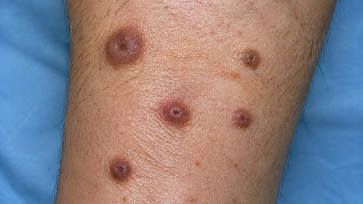
A new freckle on the collarbone, a faint brown patch across the cheek, or a sudden dark blemish on the shoulder—most of us collect marks across our skin over time. For the vast majority, these spots are merely the visible tally of years spent under daylight. For a crucial few, however, they represent an urgent call for clinical attention. The challenge lies in the fact that to the untrained eye, benign sun damage and malignant changes can look deceptively alike.
The Red Flags: When to Seek a Clinical Opinion
Before considering cosmetic fixes, the absolute priority is safety. A lesion that is actively evolving demands medical assessment. The standard clinical benchmark is the ABCDE criteria:
| Sign | Feature to Monitor | What to Look For |
| A | Asymmetry | One half of the spot fails to match the other |
| B | Border | Edges appear ragged, notched, blurred, or poorly defined |
| C | Colour | Non-uniform shades, displaying mixtures of brown, black, red, pink, or white |
| D | Diameter | Typically wider than 6mm (roughly the size of a pencil eraser), though smaller lesions can still pose risks |
| E | Evolution | Noticeable shifts over time in size, contour, elevation, pigment, or surface texture |
Beyond this checklist, pay attention to lesions that itch, crust, weep, bleed, or persistently fail to heal. Dermatologists place enormous weight on the "ugly duckling" sign—a standalone spot that simply looks and feels discordant with every other mark on your body.
With melanoma rates climbing across the UK, early detection remains the single most critical factor in successful treatment. Those with extensive mole counts, an affinity for burning easily, a history of blistering sunburns, or a family history of skin cancer should exercise a particularly low threshold for clinical review.
What Cutaneous Pigmentation Actually Is
Pigmentation is not an inherent skin flaw; it is a natural defence system. Melanocytes—the specialised cells stationed within the epidermis—generate melanin to shield cellular DNA against ultraviolet (UV) assault. When sun exposure triggers this shield, the skin freckles or tans.
When melanin production becomes dysregulated, it tends to manifest in distinct clinical presentations:

-
Solar Lentigines: Frequently termed "sun spots" or "age spots," these are well-demarcated, flat, tan-to-brown patches that crop up on persistently exposed areas, such as the face, forearms, and bald scalps.
-
Melasma: Broad, symmetrical patches with diffuse edges, commonly presenting across the forehead, bridge of the nose, and upper lip. Strongly driven by hormonal shifts—such as pregnancy, hormone replacement therapy, or oral contraceptives—it is notoriously persistent.
-
Post-Inflammatory Hyperpigmentation (PIH): Residual discolouration left behind following epidermal trauma, whether from active acne, eczema, or abrasions. PIH tends to be deeper and significantly more enduring in richer skin tones.
-
Seborrhoeic Keratoses: Harmless, waxy, slightly elevated lesions that often look as though they have been pasted onto the skin surface. While benign, their irregular texture often leads people to mistake them for suspicious moles.
The Limits of Skincare: Treating Aesthetics, Not Pathology
A common error is attempting to correct a changing, suspicious lesion with an over-the-counter brightening serum instead of securing a medical appointment. Active skincare formulations cannot eradicate or prevent cutaneous malignancies. What high-grade cosmeceuticals can accomplish is calming superficial unevenness, shielding healthy tissue, and managing benign pigmentation.
-
Broad-Spectrum Sun Protection: Without rigorous UV defence, treating discolouration is futile. A robust daily broad-spectrum SPF prevents existing melanin deposits from darkening while forestalling further photoageing. Hybrid formulas combining physical and chemical filters provide broad defence, while lightweight mist formats simplify regular reapplication throughout the day.
-
Topical Antioxidants: Daily application of a concentrated Vitamin C serum curbs the free-radical cascade induced by daylight exposure. Over time, it helps fade stubborn pigmentation while stimulating structural collagen synthesis.
-
Cellular Turnover and Exfoliation: Retinoids and chemical exfoliants (such as glycolic acid) serve as foundational agents to accelerate cell renewal, sloughing away superficial pigment and evening out the skin's texture. When complemented by botanical soothing agents, these actives minimise inflammation and reduce post-blemish staining at the source.
The Proper Order of Operations
-
Secure a Professional Diagnosis First: Arrange an appointment with your GP, a dermatologist, or a qualified skin specialist for any lesion displaying irregular behaviour or persistent change. Within the UK, you can seek an NHS referral via your GP or book an appointment through private dermatology clinics.
-
Track Objective Evolution: Maintain high-resolution, well-lit, dated photographs of any borderline marks. Relying purely on memory makes gradual changes difficult to pinpoint.
-
Establish a Measured Routine: Once a lesion has received medical clearance, focus on consistent, targeted skincare. Long-term adherence to daily sun protection, topical antioxidants, and gentle exfoliation will deliver far greater results over twelve months than any rapid-fix cosmetic claim.
Skin cancer remains highly treatable when caught early, but no cosmetic bottle can make a dangerous lesion safe. Have the suspicious marks reviewed first; establish your skincare regime after