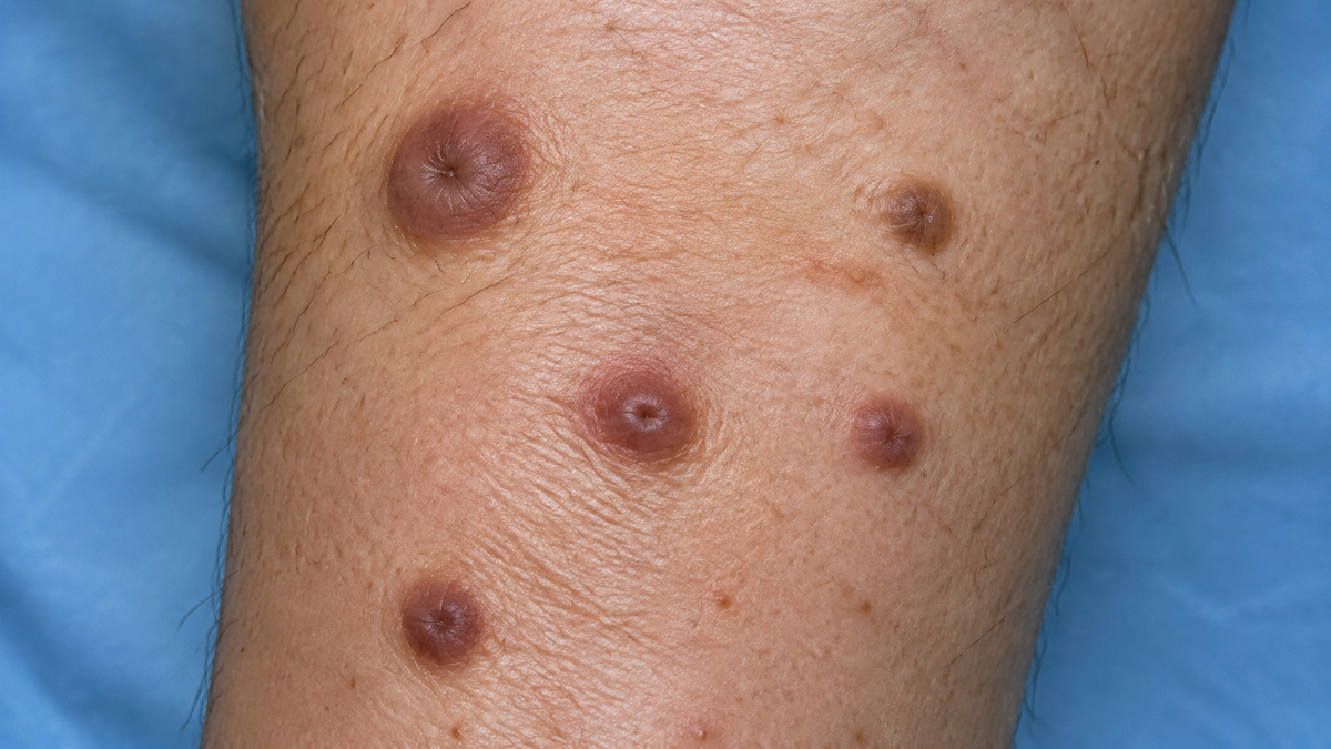
Understanding Dermatofibromas: From Clinical Appearance to Microscopic Detail
Although dermatofibromas are entirely harmless for the vast majority of individuals, they remain one of the most frequent reasons for a patient to visit a GP or a specialist dermatology clinic. These small, firm nodules occur commonly on the lower extremities. The principal challenge they present is not the threat posed by the lump itself, but rather the diagnostic dilemma it creates. Because a benign dermatofibroma can easily be mistaken for a malignant melanoma, a dermatofibrosarcoma protuberans, or various other serious soft-tissue tumours, medical practitioners naturally adopt a cautious approach whenever uncertainty arises.
By looking closely at how these lesions form, what a dermatoscope reveals, and how histopathologists identify them, it becomes clear why such an inconspicuous bump receives significant medical attention.
What Is a Dermatofibroma?
A dermatofibroma—also referred to as a fibrous histiocytoma—is a non-cancerous growth composed of fibrous tissue within the dermis, the skin’s deeper structural layer. They are widely believed to develop following minor localized trauma, such as an insect bite, a thorn prick, an ingrown hair, or a small shaving cut. Instead of healing normally, the tissue undergoes an exaggerated inflammatory and fibrotic response, creating a small, persistent nodule.
-
Key Physical Features:
-
Typically measure between $5\text{ mm}$ and $10\text{ mm}$ across.
-
Feel firm and fixed to the underlying dermal structures, preventing them from sliding freely beneath the skin.
-
Range in colour from pinkish-tan and reddish-brown to dark chocolate brown, often featuring a darker rim with a lighter central zone.
-
-
The "Pinch Test" (Dimple Sign): The most reliable clinical test at the bedside is the dimple sign. When gently compressed between the index finger and thumb, the centre of the lesion sinks inward instead of pushing outward,because the internal fibrous strand anchors the surface to deeper tissues.
-
Demographics: While typically appearing as single, isolated lumps, multiple lesions can occur. They affect individuals of all ages—peaking in early-to-middle adulthood—and are more frequently observed in women, largely owing to their predilection for the lower legs.
Why Accurate Diagnosis Is Vital
Dermatofibromas are clinical chameleons, capable of mimicking several far more serious skin conditions. A firm,pigmented nodule on an adult's leg may prove to be a benign dermatofibroma, but visually it can mimic:
-
Nodular Melanoma: The most aggressive melanoma variant, which tends to grow thick rapidly and carries a high risk if missed.
-
Dermatofibrosarcoma Protuberans (DFSP): A locally invasive, slow-growing malignant tumour that early on looks deceptively benign and demands broad surgical removal.
-
Other Mimics: Blue naevi, Spitz naevi, keloid scars, or cutaneous metastases.
While a dermatofibroma generally stays unchanged over many months or years, a melanoma typically displays noticeable progression. However, patient history alone cannot confirm a diagnosis. Because missing a malignant growth carries severe consequences, doctors rely on dermoscopy and tissue biopsies rather than visual inspection with the naked eye.
Dermoscopic Examination and Triage
Dermoscopy involves inspecting skin lesions using a hand-held light source equipped with high magnification and cross-polarised filters to eliminate surface glare. This allows clinicians to observe underlying dermal and epidermal architecture.
       [ Outer Edge ]  -->  Faint, delicate brown pigment ring / network
             |
       [ Central Area ] -->  Scar-like white patch (dermal fibrosis & dense collagen)
             |
       [ Periphery ]   -->  Fine, linear or branching blood vessels
When these features appear together, they create a characteristic "signature" that strongly supports a diagnosis of dermatofibroma.
This examination serves as a crucial triage step:
-
Classic Pattern: If the classic central white patch and fine peripheral network are present, the practitioner can reassure the patient and avoid unnecessary surgical procedures.
-
Atypical Pattern: If irregular pigmentation, broad network patterns, blue-grey veiling, or asymmetrical structures are detected, a biopsy is promptly performed to rule out malignancy.
Microscopic Analysis (Histopathology)
When a definitive answer is required, a tissue sample is evaluated under a microscope. A mature dermatofibroma is characterized by a proliferation of spindle-shaped fibroblasts and myofibroblasts forming short, interlacing bundles within the dermis. Dense collagen bundles wrap around these cells, frequently arranged in a distinct whorled or cartwheel-like (storiform) pattern.
-
Epidermal Changes: The skin layer directly above the nodule often displays thickening (acanthosis) and a central depression that corresponds directly to the dimple sign seen during physical examination.
-
Histological Variants: Pathologists recognise several subtypes, including cellular, aneurysmal (which contains blood-filled spaces), atrophic, and haemosiderotic variants (which contain iron deposits from past bleeding and closely resemble melanoma).
-
Immunohistochemistry: Pathologists use specific antibody stains—such as factor XIIIa and CD34—to reliably differentiate complex dermatofibroma variants from aggressive conditions like dermatofibrosarcoma protuberans.
Management and Patient Care
In the vast majority of cases, reassurance is all that is required. Confirmed dermatofibromas can be safely left alone, and they frequently fade or become less prominent over time.
| Scenario | Action / Considerations |
| Asymptomatic Lesions | No intervention required; monitor for changes. |
| Symptomatic or Irritated | Surgical excision or shave removal can be considered if the lump catches on clothing or becomes inflamed. |
| Cosmetic Concerns | Patients should be advised that surgical removal leaves a permanent scar that may appear more noticeable than the initial lesion. |
| Alternative Therapies | Treatments like cryotherapy or laser therapy may flatten the surface but rarely eradicate the fibrous root entirely. |
Key Patient Advice
Any skin lesion that exhibits recent changes—such as rapid growth, bleeding, ulceration, color alteration, or new pain—should be assessed promptly by a healthcare professional. Self-diagnosis using online imagery is unreliable and risks delaying critical medical care. Ultimately, the integration of careful physical examination, dermoscopy, and histopathology ensures that diagnostic uncertainty is safely replaced with clinical certainty.