
Basal Cell Carcinoma (BCC): Symptoms, Causes & UK Treatments
Key changes include updating to British spelling (e.g., carcinoma, tumour, localised), swapping US medical terminology for UK equivalents (e.g., GP, dermatologist, sunbed), and aligning healthcare pathways with the National Health Service (NHS) framework.
Basal Cell Carcinoma (BCC): Overview, Symptoms, and Treatment
Basal cell carcinoma, often shortened to BCC, is the most common type of skin cancer. It begins in the basal cells, which are found in the deepest layer of the epidermis—the outermost layer of the skin. These cells are responsible for producing new skin cells as old ones shed. When DNA damage causes basal cells to grow uncontrollably, basal cell carcinoma can develop.
Although the word “cancer” can sound frightening, basal cell carcinoma is usually slow-growing and rarely spreads to distant parts of the body. However, it should not be ignored. If left untreated, BCC can grow deeper into the skin and damage nearby tissue, nerves, cartilage, or bone. Early detection and treatment are the best ways to prevent complications and achieve excellent outcomes.
This article explains what basal cell carcinoma is, what causes it, how to recognise it, how it is diagnosed and treated, and what you can do to reduce your risk.
What Is Basal Cell Carcinoma?

Basal cell carcinoma is a form of non-melanoma skin cancer. It develops from basal cells in the skin and most often appears on areas frequently exposed to the sun, such as the face, scalp, ears, neck, shoulders, arms, and hands. It can also appear on parts of the body that receive little sun exposure, though this is less common.
BCC is strongly linked to ultraviolet (UV) radiation, especially from sunlight and sunbeds. Over time, UV radiation can damage the DNA inside skin cells. When the body cannot repair this damage properly, abnormal cells may begin multiplying.
Basal cell carcinoma is generally considered less aggressive than melanoma, but it can still be locally destructive. This means it may invade surrounding tissue if it grows for a long time. Because many BCCs develop on the face, untreated lesions can lead to cosmetic and functional problems, especially around the nose, eyelids, lips, and ears.
Who Is at Risk?
Anyone can develop basal cell carcinoma, but some people have a higher risk than others. Risk factors include:
-
Frequent sun exposure: People who spend a lot of time outdoors for work or recreation have an increased risk.
-
History of sunburns: Severe or repeated sunburns, especially during childhood, can raise the chance of developing skin cancer later in life.
-
Use of sunbeds: Indoor tanning exposes skin to concentrated, high-intensity UV radiation.
-
Fair skin: People with light skin, light-coloured eyes, blonde or red hair, and freckles are more vulnerable to UV damage.
-
Older age: BCC becomes more common with age because sun damage accumulates over time.
-
Personal history of skin cancer: Having had one BCC increases the risk of developing another.
-
Family history: Genetics can play a role in skin cancer susceptibility.
-
Weakened immune system: People taking immunosuppressive medications (such as organ transplant recipients) or living with immune-suppressing conditions have a higher risk.
-
Prior radiation therapy: Previous radiotherapy treatments may increase the risk of skin cancer in the treated areas.
-
Certain genetic conditions: Rare disorders, such as Gorlin syndrome (basal cell naevus syndrome), can cause multiple BCCs.
While basal cell carcinoma is more common in fair-skinned individuals, it can occur in people of all skin tones. In darker skin, skin cancers may be diagnosed later because they are less expected or harder to notice. Any new, changing, bleeding, or non-healing skin lesion deserves medical attention.
Common Signs and Symptoms
Basal cell carcinoma can look different from person to person. It may be mistaken for a pimple, scar, eczema patch, or a sore that will not heal. Because it often grows slowly, people may ignore it for months or even years.

Common signs include:
-
A pearly or shiny bump: This may be pink, red, white, skin-coloured, brown, or black.
-
Visible tiny blood vessels: Some BCCs have small red or purple vessels (telangiectasia) spidering across the surface.
-
A sore that does not heal: It may bleed, crust over, heal temporarily, and then reopen.
-
A flat, scaly patch: This may look like dry skin, psoriasis, or dermatitis.
-
A scar-like area: Some BCCs appear waxy, pale, firm, or slightly indented.
-
A bleeding or easily irritated spot: The lesion may bleed after washing, shaving, or minor rubbing.
-
A raised growth with rolled edges: Some BCCs develop a central dip or ulcer.
Not every basal cell carcinoma looks “classic”. Pigmented BCCs, for example, can appear brown, blue, or black and may resemble melanoma. Any suspicious spot should be checked by a healthcare professional, especially if it is changing, growing, bleeding, painful, or not healing.
Types of Basal Cell Carcinoma
Doctors and pathologists classify BCC into several subtypes based on how it looks clinically and under a microscope. The subtype can affect your NHS treatment options.
Nodular Basal Cell Carcinoma
This is the most common form. It often appears as a shiny, pearly bump, usually on the face. It may have visible blood vessels and can bleed or ulcerate easily.
Superficial Basal Cell Carcinoma
This type often appears as a red, scaly patch, commonly on the torso (trunk), shoulders, arms, or legs. It may be mistaken for eczema or psoriasis. Superficial BCC may be suitable for non-surgical topical treatments in selected cases.
Morpheaform or Infiltrative Basal Cell Carcinoma
This subtype tends to appear as a firm, scar-like area. It can be more difficult to see clearly because its edges blend into normal skin. It may grow deeper and wider than expected, so it usually requires precise surgical removal.
Pigmented Basal Cell Carcinoma
Pigmented BCC contains dark melanin and may look brown, black, or blue. Because it can closely resemble melanoma, proper evaluation and a biopsy are crucial.
How Basal Cell Carcinoma Is Diagnosed
Diagnosis usually begins with an examination by a General Practitioner (GP). If they suspect a skin cancer, you will be referred to a specialist, typically a dermatologist or a plastic surgeon, via the NHS referral pathway.
The specialist will look closely at the spot using a dermatoscope—a handheld tool that magnifies the skin and lights up the deeper structures to reveal patterns not visible to the naked eye.
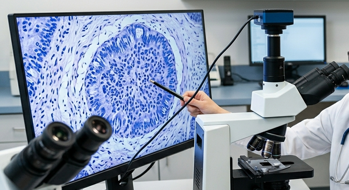
If basal cell carcinoma is suspected, the specialist will usually perform a skin biopsy. This involves numbing the area with a local anaesthetic, removing a small sample of the lesion (or sometimes the entire lesion), and sending it to a laboratory. A pathologist examines the tissue under a microscope to confirm whether cancer cells are present and to identify the subtype.
Common biopsy methods include:
-
Shave biopsy: A thin layer of tissue is shaved off from the surface.
-
Punch biopsy: A small, circular tool removes a deeper core sample.
-
Excisional biopsy: The entire suspicious area is cut out.
A biopsy is important because different skin conditions and cancers can look very similar. Treatment planning depends entirely on the diagnosis, size, location, depth, and microscopic features of the tumour.
Treatment Options
Basal cell carcinoma is highly treatable, especially when caught early. In the UK, your treatment plan will depend on the tumour’s size, location, subtype, and whether it is a recurrence, alongside your overall health.
Surgical Excision
In a standard surgical excision, the doctor cuts out the cancer along with a safety margin of normal-looking skin. The wound is closed with stitches, and the tissue is sent to a lab to ensure the margins are completely clear of cancer. This method is widely used and has a very high cure rate.
Mohs Micrographic Surgery
Mohs surgery is a specialised technique often used for BCCs on the face, ears, scalp, or other areas where preserving healthy skin is vital for appearance and function. During Mohs surgery, the consultant removes the cancer layer by layer, immediately examining each layer under a microscope while you wait. This continues until no cancer cells remain.
Note: Due to its specialised nature, Mohs surgery on the NHS is typically reserved for high-risk zones (like around the eyes or nose) or recurrent tumours.
Curettage and Cautery (Electrodessication)
This method involves scraping away the soft tumour tissue with a spoon-shaped instrument (a curette) and using an electrical current to stop bleeding and destroy any remaining cancer cells. It is highly effective for certain small, low-risk BCCs on the body or limbs.
Cryotherapy
Cryotherapy uses extreme cold, usually liquid nitrogen, to freeze and destroy abnormal cells. It may be considered for small, superficial lesions, but it is not appropriate for deeper or more aggressive BCCs.
Topical Medications
Some superficial BCCs can be treated at home with prescription creams, such as imiquimod (Aldara) or 5-fluorouracil (Efudix). These treatments trigger the immune system or target cancer cells directly over several weeks and require careful follow-up by your clinical team.
Photodynamic Therapy (PDT)
Photodynamic therapy involves applying a light-sensitive cream to the skin, waiting a few hours for it to be absorbed by the abnormal cells, and then activating it with a special wavelength of light. This destroys the cancerous cells while leaving healthy tissue relatively undamaged. It is commonly used for superficial BCCs.
Radiotherapy
Radiation therapy may be used when surgery is not possible or preferred—particularly in older adults or individuals with health conditions that make minor surgery difficult. It can also be useful for tumours in anatomically challenging locations.
Targeted Therapy and Immunotherapy
For rare, advanced cases where the BCC has grown exceptionally deep into surrounding structures or has spread (metastased), systemic medications may be used. These include targeted drugs that block the hedgehog signalling pathway, such as vismodegib, or specific immunotherapies.
What Happens If BCC Is Left Untreated?
Basal cell carcinoma usually grows slowly, but it will not simply disappear. Without medical intervention, it will continue to enlarge and invade nearby structures. On the face, this can lead to significant tissue damage. For example, a BCC near the nose, eyelid, or ear can erode into cartilage or bone and interfere with normal functions like vision or breathing.
Larger, neglected tumours require much more extensive surgery to remove, which can leave more noticeable scars and require complex reconstructive surgery or skin grafts. Early treatment is always simpler, less invasive, and yields the best cosmetic result.
Prevention: How to Reduce Your Risk
The most effective prevention strategy is protecting your skin from UV damage. This does not mean avoiding the outdoors entirely, but practicing safe sun habits.
-
Use sunscreen daily: Choose a broad-spectrum sunscreen with a Sun Protection Factor (SPF) of 30 or higher, and an UVA rating of 4 or 5 stars. Apply it generously and reapply every two hours when outdoors, or immediately after swimming or sweating.
-
Wear protective clothing: Opt for tightly woven long sleeves, wide-brimmed hats, and UV-blocking sunglasses.
-
Seek shade: Keep out of the sun when UV rays are strongest, typically between 11 am and 3 pm from March to October in the UK.
-
Avoid sunbeds: Indoor tanning exposes skin to dangerous levels of UV radiation, drastically increasing skin cancer risk and accelerating skin ageing.
-
Check your skin regularly: Conduct monthly self-exams to check for new, changing, bleeding, or non-healing spots.
-
Attend follow-ups: If you have a history of skin cancer, ensure you attend any scheduled monitoring appointments with your care team.
Sun protection should start early in life, but it is never too late to benefit. Even after years of sun exposure, protecting your skin now will prevent further damage and lower your risk of future skin cancers.
Living After a Basal Cell Carcinoma Diagnosis
Receiving a BCC diagnosis can cause anxiety, and it is entirely normal to worry about recurrence, scarring, or future skin cancers. Because having one basal cell carcinoma increases your risk of developing another, proactive follow-up care is essential.
After your treatment, your GP or dermatologist will advise you on what to look out for. You should monitor the treated area carefully for signs of recurrence—such as a new bump, persistent redness, or a sore that reopens.
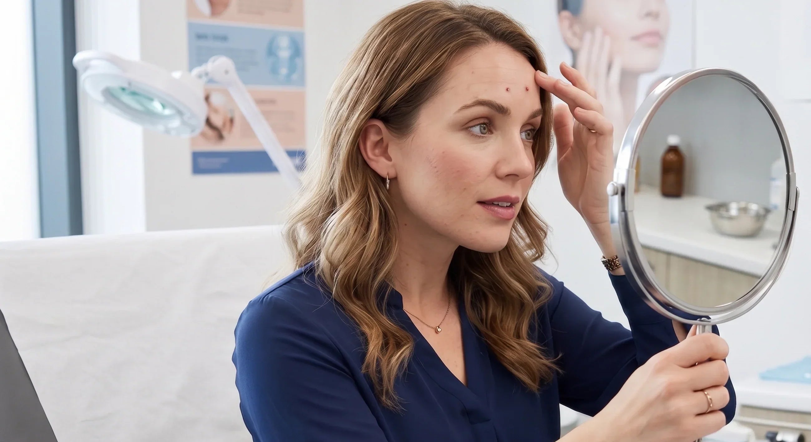
Get to know your skin. Use a full-length mirror, a hand mirror, or ask a partner or close friend to check hard-to-see areas like your back, scalp, and behind your ears.
When to See a Doctor
You should make an appointment with your GP if you notice:
-
A sore, ulcer, or scab that does not heal within 4 weeks.
-
A spot that bleeds, oozes, crusts, or repeatedly opens.
-
A shiny, pearly, or translucent bump.
-
A flat, scaly red patch that persists despite moisturising.
-
A scar-like area that appears out of nowhere without a prior injury.
-
A mole or dark spot that changes in size, shape, or colour.
Your GP can evaluate the spot and, if necessary, fast-track a referral to a specialist clinic. Early evaluation makes treatment straightforward and highly successful.
Conclusion
Basal cell carcinoma is the most common skin cancer, but it is also one of the most treatable when detected early. It develops from basal cells in the skin and is most often linked to UV exposure from sunlight or sunbeds. While BCC rarely spreads to distant organs, it can grow into nearby tissue and cause significant damage if ignored.
Knowing the warning signs—such as a non-healing sore, shiny bump, scaly patch, or bleeding spot—can help you seek care promptly. Diagnosis usually requires a biopsy, and NHS treatment options range from standard surgical removal and Mohs surgery to topical creams, photodynamic therapy, and radiotherapy.
The best defense is a combination of sun protection, regular skin checks, and timely medical care. Protecting your skin today will reduce your risk of future skin cancers and help keep your skin healthy for years to come.

,%20swapping%20US%20medical%20terminology%20for%20UK%20equivalents%20(e.g.,%20GP,%20dermatologist,%20sunbed),%20and%20aligning%20healthcare...)