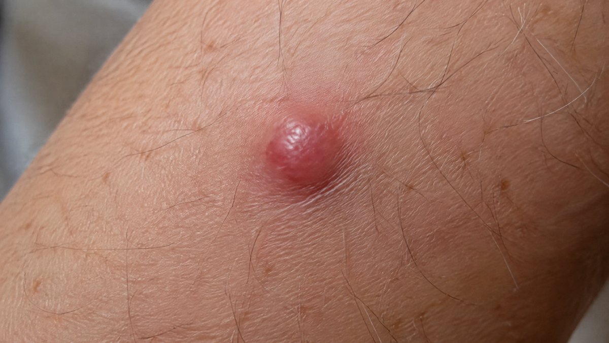
Article: 5 Mole Warning Signs You Shouldn't Ignore | Melanoma Guide
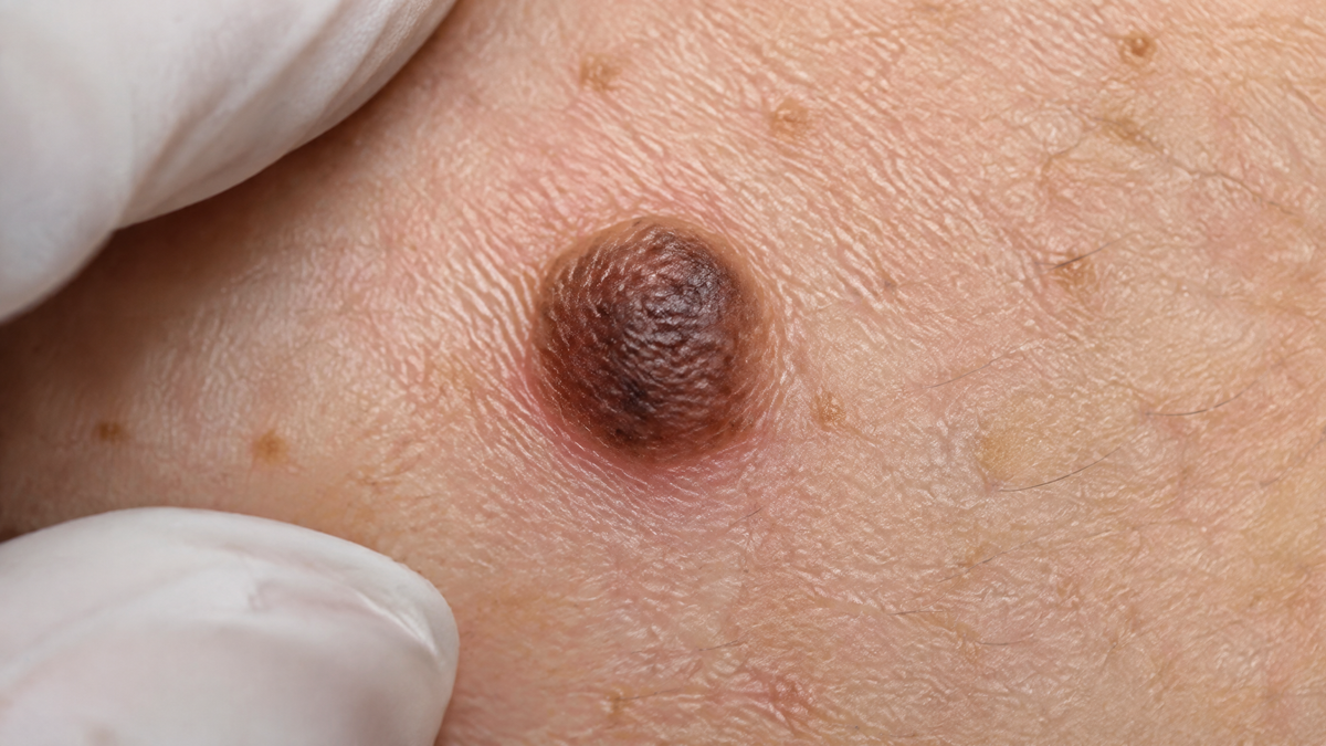
5 Mole Warning Signs You Shouldn't Ignore | Melanoma Guide
When to Get a Mole Checked: Five Warning Signs You Should Never Ignore
Nearly all of us have moles somewhere on our bodies — whether it is a small speck across the collarbone, a childhood beauty mark, or a cluster of freckles on the shoulders. In almost every instance, these are simply benign collections of melanocytes quietly coexisting with us throughout our lives. However, a small minority undergo alterations, and catching those changes early is the single most critical factor in treating skin cancer successfully.
Melanoma rates across the United Kingdom, Australia, and New Zealand have climbed substantially over recent decades. While public awareness often associates sun risk with summer holidays abroad, routine UV exposure at home presents just as much of an accumulative danger. Crucially, early-stage melanoma is one of the most curable malignancies known to medicine. The difficulty is that it rarely aches, stings, or bleeds in its initial phase; it develops quietly, making vigilance your most valuable safeguard.
Five Key Indicators That Warrant a Doctor’s Evaluation
1. Changes in Dimensions, Contour, or Pigmentation
Evolution is the vital red flag. A spot that has noticeably expanded beyond the size of a pencil eraser, or one with blurred, notched, or jagged edges where it was once neatly defined, demands a clinical review. The same applies to shade: normal moles tend to be a uniform brown or tan, whereas concerning lesions often display mixed patches of dark brown, black, pink, slate-grey, or white.
Dermatologists routinely teach the ABCDE framework to identify these shifts:
-
A — Asymmetry: One half does not match the other.
-
B — Border: Edges are irregular, notched, or poorly defined.
-
C — Colour: Multiple shades or uneven distribution of pigment.
-
D — Diameter: Measuring across by more than 6mm.
-
E — Evolving: Any noticeable transition in appearance over weeks or months.
Using your smartphone to photograph existing marks under consistent lighting creates a reliable baseline. If you look back six months later and spot clear progression, arrange an appointment rather than adopting a wait-and-see mindset.
2. The "Ugly Duckling" That Breaks the Mould
Most individuals possess moles that share a consistent personal pattern in terms of size, shape, and tone. Dermatologists frequently rely on the "ugly duckling" concept: a solitary lesion that looks entirely disparate from the surrounding spots on your torso or limbs should be viewed with suspicion, even if it does not neatly breach every ABCDE criterion. Comparing bilateral areas — your left arm against your right, or your upper legs — makes an outlier far easier to spot.
3. Unexplained Irritation, Crusting, or Bleeding
A standard mole should remain symptom-free. If a lesion becomes persistently itchy over several weeks, develops an unhealing crust, or weeps blood after minor contact with a bath towel, your body is signalling a problem. While everyday friction from collars, waistbands, or razors can cause superficial irritation, a recurrently weeping sore that lingers beyond three to four weeks requires a definitive assessment by a doctor rather than another plaster.
4. The Emergence of Fresh Lesions After 30
Children, teenagers, and young adults naturally produce new moles as their bodies mature. By the time you enter your thirties, however, new pigment deposits should slow markedly. A completely novel dark mark appearing in mid-to-late adulthood — particularly one that grows quickly over a few months — deserves prompt clinical scrutiny.
Your threshold for booking an assessment should be significantly lower if you fall into an elevated risk category:
-
A personal or first-degree family history of skin cancer
-
Over 50 to 100 moles across your body
-
Pale skin that burns or freckles easily in the sun
-
A history of blistering sunburns during childhood
5. Moles Tucked Away in Unseen Locations
Lesions hidden on the scalp, behind the ears, across the lower back, between the toes, on the soles of the feet, or beneath the nail beds are frequently detected late simply because they evade everyday view. This delayed diagnosis carries genuine risk; acral lentiginous melanoma, which targets the palms, soles, and nail beds, accounts for a higher proportion of cases in individuals with darker skin tones and is often caught late.
Make a monthly self-inspection routine standard practice. Enlist a partner or family member to inspect your scalp and back, and use a hand mirror to check the bottoms of your feet. Keep an eye out for any unprovoked, longitudinal dark streaks running beneath a fingernail or toenail.
What to Expect During an Appointment and Removal
A consultation typically begins with your GP or a private dermatologist carrying out an examination using a dermatoscope — a handheld instrument combining high magnification with specialised lighting that illuminates underlying dermal structures invisible to the naked eye. In an increasing number of NHS and private clinics, clinicians also draw on validated digital imaging and AI-assisted analysis tools to benchmark suspicious lesions against vast comparative datasets.

If an anomaly is identified, you will undergo a minor procedure rather than immediate major surgery:
[Clinical Assessment via Dermatoscope]
                   │
                   ▼
[Excisional Biopsy under Local Anaesthetic (15–30 mins)]
                   │
                   ▼
[Histopathology Results Received (1–2 weeks)]
        ┌──────────┴──────────┐
        ▼                     ▼
[Benign / Clear]       [Melanoma Confirmed]
                       - Wide Local Excision
                       - Staging (Depth Assessment)
                       - Lymph Node Review if required
The gold standard for a concerning mole is a diagnostic excision carried out in a clinic room under local anaesthetic. The doctor injects an anaesthetic agent — which pinches for a few seconds before the area goes entirely numb — removes the lesion alongside a slender safety margin of healthy tissue, and secures the wound with a few small sutures. These stitches are routinely removed after 7 to 14 days, with the tissue sent straight to a pathology laboratory for microscopic evaluation.
Should the histopathology report confirm a malignancy, next steps are governed entirely by the tumour's depth (the Breslow thickness, measured in fractions of a millimetre). Superficial melanomas generally require nothing more than a secondary wider local excision to guarantee clear margins. Deeper or more advanced lesions may involve a sentinel lymph node biopsy, followed by targeted therapies or immunotherapy — modern systemic treatments that have radically improved prognosis over the past decade.
Understanding the Odds: Action Over Anxiety
The overwhelming majority of skin assessments end with reassurance: the vast majority of excised or surveyed lesions turn out to be completely benign. However, pursuing a quick professional opinion is about stacking the odds in your favour.
When melanoma is identified at Stage 1, the UK five-year survival rate stands above 95 per cent. That figure drops considerably once the disease spreads to distant organs, which is precisely why lingering doubt must never be brushed aside. Doctors would invariably prefer to review twenty harmless, healthy moles than see a single late-stage tumour that could have been resolved two years earlier.
Sensible Prevention for the Long Term
Taking care of your skin does not necessitate hiding indoors; it simply demands pragmatic habits:
-
Apply broad-spectrum sunscreen with an SPF of at least 30 and high UVA star protection, reapplying every two hours when outdoors.
-
Rely on wide-brimmed hats, UV-blocking sunglasses, and tightly woven clothing during peak midday hours.
-
Keep children thoroughly shielded from intense ultraviolet rays, as severe childhood sunburns significantly drive up adult melanoma rates.
-
Avoid sunbeds altogether; an artificial ultraviolet tan reflects cellular DNA damage, not vitality.
This guide provides general health information and does not replace dedicated clinical judgement. If you notice a mole that is changing, atypical, or persistently irritating, book an examination with your GP or a registered dermatologist for an accurate diagnosis.