Article: Red Lump on Your Hand? Causes, Red Flags & When to See a GP
Red Lump on Your Hand? Causes, Red Flags & When to See a GP
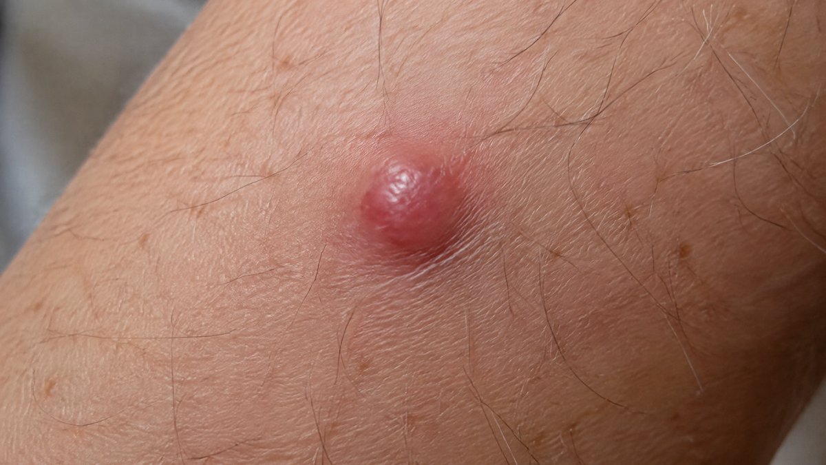
A sudden red lump on the hand is a surprisingly unsettling sight. Whether it flared up following a mild scuff against a doorframe or emerged quietly across a knuckle without warning, waiting to see what happens is a common first reaction. While the vast majority of hand lesions are entirely benign, delaying review is also why significant skin conditions are occasionally caught later than they ought to be.
Because the hands are in perpetual motion, frequently knocked, heavily exposed to weather, and constantly in your line of sight, every subtle change is immediately apparent.
Red Flags: When to Seek Immediate Medical Review
Before reviewing potential diagnoses, note the warning signs that warrant prompt evaluation by a GP or dermatologist. Arrange an appointment if you identify any of the following:
-
Rapid expansion: A lump that noticeably grows over weeks rather than months.
-
Spontaneous bleeding: Lesions that bleed without significant trauma, ooze recurrently, or prove difficult to stem.
-
Non-healing ulceration: Any broken skin or cratering that fails to resolve within four weeks (a key criterion for urgent NHS suspected cancer pathways).
-
Evolution of an existing mark: A longstanding blemish that begins itching, bleeding, altering shape, or changing shade.
-
Irregular margins and asymmetry: Outlines that appear jagged, notched, or unevenly weighted.
-
Variegated pigmentation: Multiple colours within the same spot (blends of red, blue, dark brown, black, or greyish-white).
-
Fixation: A lump anchored firmly to the underlying tissue or tendon sheath rather than sliding freely beneath the skin.
-
Associated systemic symptoms: Unexplained fever, drenching night sweats, fatigue, or unintended weight loss.
The classic ABCDE framework (Asymmetry, Border, Colour, Diameter, Evolving) remains vital. For nodules, E (Evolution) is paramount — any growth that refuses to settle or is actively changing requires professional assessment.
What Defines a True Skin Nodule?
In clinical dermatology, a nodule is a solid, raised lesion distinct from a superficial spot (papule) by virtue of its depth and dimension. Typically measuring between 5mm and 10mm or greater, a nodule extends into the deeper dermis or subcutaneous layer, meaning it has palpable volume beneath the skin surface rather than merely sitting on top.
Colour provides further diagnostic clues:
-
Blanching lesions: When pressure is applied and the redness temporarily fades to pale before refilling with blood, an underlying vascular issue is typical.
-
Non-blanching lesions: Firm nodules that do not pale under pressure frequently suggest fibrous overgrowths, cellular infiltration, inflammatory deposits, or neoplastic changes.
Common Causes of Red Hand Lumps
| Condition | Typical Appearance & Behaviour | Clinical Significance |
| Pyogenic Granuloma (Lobular capillary haemangioma) | Rapidly enlarging, bright red, glossy, fleshy vascular bump. Prone to profuse bleeding from minor knocks. | Entirely benign, yet can closely mimic amelanotic melanoma or squamous cell carcinoma; excision/cautery is standard. |
| Dermatofibroma | Firm, reddish-brown or dusky pink nodule, frequently following insect stings or minor trauma. | Displays the "dimple sign" (puckers inward when pinched). Completely harmless. |
| Cherry Angioma | Small, distinct cherry-red or purple vascular spot, stable over years. | Benign vascular collection; requires intervention only if repeatedly irritated or catching on sleeves. |
| Chilblains (Perniosis) | Pruritic, tender, purplish-red inflammatory swellings triggered by damp, cold British weather. | Usually transient; recurrent or persistent episodes warrant testing for underlying autoimmune disorders (e.g. lupus). |
| Gouty Tophi & Rheumatoid Nodules | Dense, chalky or flesh-toned to erythematous lumps positioned over joints, knuckles, and tendon paths. | Secondary to systemic joint disease (monosodium urate deposition or severe rheumatoid arthritis). |
| Cutaneous Infections | Hot, throbbing, erythematous swelling, occasionally accompanied by pus or flu-like symptoms. | Abscesses or infected epidermoid cysts requiring drainage or antibiotic intervention. |
| Squamous Cell Carcinoma (SCC) & Keratoacanthoma | Rapidly developing, hyperkeratotic nodule often featuring a central keratin crater or raised borders on sun-exposed skin. | Malignant skin cancer linked to cumulative ultraviolet radiation; requires prompt surgical management. |
| Amelanotic Melanoma | Firm, non-pigmented, pinkish-red nodule lacking the classic dark brown or black melanin. | Dangerous melanoma subtype that disguises itself as an innocent inflammatory papule or vascular lump. |
What to Expect During a Clinical Consultation
Attending a skin clinic is straightforward and non-invasive:
-
Clinical History: The clinician will ask when the lump first presented, whether it bleeds or aches, any history of outdoor UV exposure, occupational hazards, and personal or family histories of skin disease.
-
Dermoscopic Examination: Using a dermatoscope (a high-resolution skin microscope using polarised illumination), the practitioner can examine deep vascular patterns and pigment structures imperceptible to the naked eye.
-
Diagnostic Biopsy: If diagnostic uncertainty persists, a minor procedure under local anaesthetic (shave, punch, or full surgical excision) will be performed. The tissue is sent for histopathological analysis, providing an absolute and definitive diagnosis.
Managing Your Skin While Awaiting Review
Immediate Self-Care Rules
-
Create a visual timeline: Photograph the nodule in bright, natural light alongside a coin or metric ruler. Repeat every fortnight to supply your clinician with clear objective evidence of growth.
-
Refrain from interference: Do not squeeze, puncture, pick, or attempt home freezing kits, drawing salves, or high-strength chemical exfoliants, which inflame the area and distort diagnostic histology.
-
Control acute bleeding: For high-flow vascular bleeding, apply firm, direct, uninterrupted pressure with a clean lint-free pad for 15 to 20 minutes without lifting to check.
The Role of Supportive Skincare Topical skincare products cannot treat or diagnose a suspicious nodule. Their legitimate function is to reinforce barrier integrity across the surrounding, weather-beaten hand tissue:

-
Barrier Restoration: Frequent handwashing and cold air deplete natural epidermal lipids. Non-greasy emollients formulated with ceramides, panthenol (Pro-Vitamin B5), or physiological lipids support moisture retention and reduce painful cracking around unaffected skin.
-
Daily UV Shielding: Because cumulative solar radiation drives squamous cell carcinoma and lentigo maligna on the dorsal hands, daily broad-spectrum SPF 30 to SPF 50+ mineral or chemical sun protection should be extended to the backs of the hands year-round.
-
Lesion Precaution: Keep active, broken, bleeding, or ulcerated nodules free of cosmetic creams, fragranced products, and serums until clinically evaluated.