Article: Can You Diagnose Skin Conditions From a Photo? | UK Guide
Can You Diagnose Skin Conditions From a Photo? | UK Guide
Can you diagnose a skin condition simply by taking a photograph? The short answer is: occasionally in part, but never with the clinical certainty required for safe medical care. While smartphone photography and digital imaging have advanced rapidly, a critical gap remains between recognising that an image resembles a known condition and receiving an accurate diagnosis with an appropriate treatment plan. Misunderstanding this distinction is precisely where patients risk their health.
The Reality of a Clinical Diagnosis
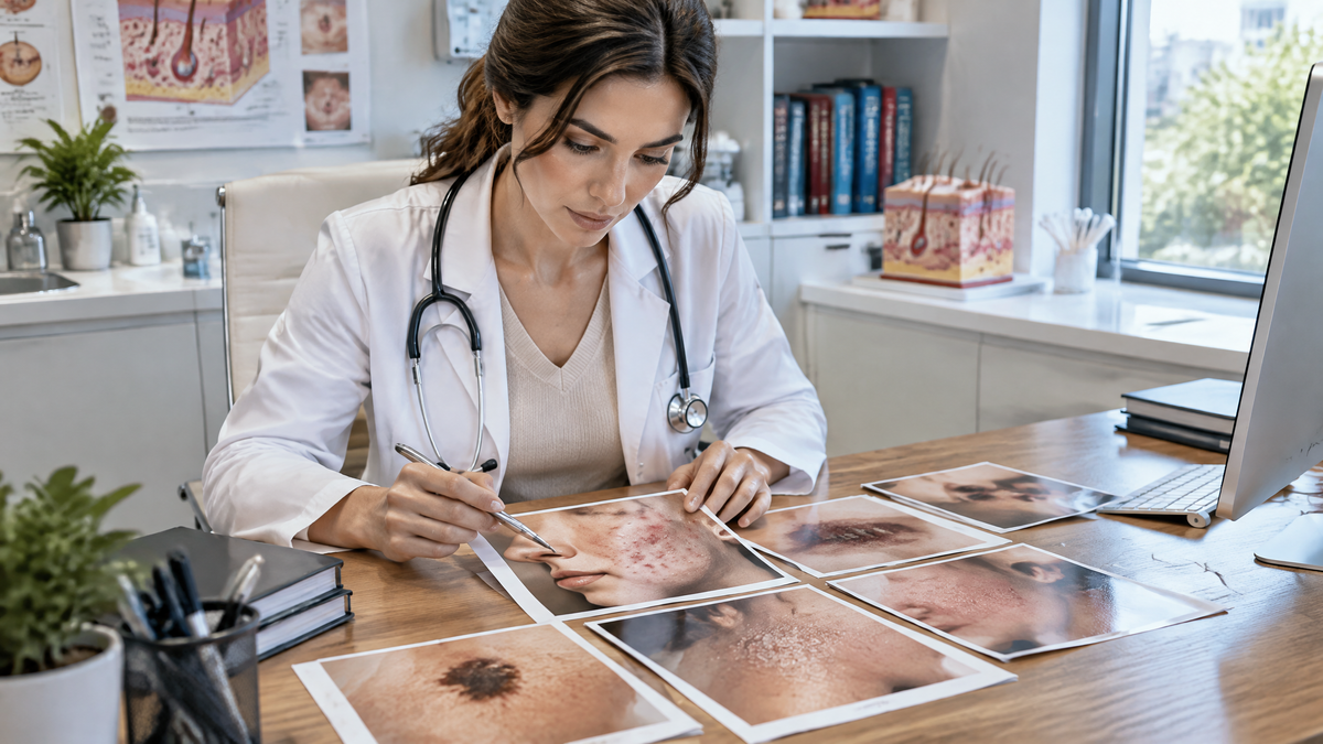
To appreciate why snapshots fall short, consider what a formal dermatological assessment actually involves. A qualified clinician gathers far more than just visual data:
-
Multisensory Examination: Beyond assessing surface colour and contour, a doctor palpates the skin to evaluate firmness, elevation, mobility, and warmth, testing whether the lesion blanches when pressed.
-
Specialised Optics: Practitioners rely on a dermatoscope—a high-magnification device utilising polarised light—to inspect subsurface structures invisible to a standard camera lens.
-
Comprehensive Review: A single spot must be contextualised alongside the rest of your skin, scalp, nails, mucous membranes, and lymph nodes.
-
Clinical History: Crucial details—such as duration, evolving sensations (itching, pain, tenderness), past medical history, family genetics, sun exposure, and prescription medications—are weighed carefully.
-
Histopathology: When ambiguity persists, a definitive diagnosis is often confirmed only after a biopsy, where tissue is examined microscopically by a pathologist.
A flat, two-dimensional mobile photo strips away almost all of this depth. Camera sensors and onboard software routinely distort true colours, struggle with subtle inflammation in darker skin tones, and obscure both scale and perspective.
Where Photo-Matching and Diagnostic Apps Fail
Treating an image search or a consumer application as a diagnostic tool creates serious clinical hazards:
| Pitfall | Why It Happens | Clinical Risk |
| Symptom Overlap | Conditions such as eczema, psoriasis, ringworm, and contact dermatitis often present as near-identical scaly red patches. | Inappropriate self-treatment can aggravate the condition. |
| Deceptive Mimicry | A harmless seborrhoeic keratosis can mimic a melanoma; early basal cell carcinomas often resemble simple stubborn spots. | Life-threatening conditions may be ignored while benign marks cause panic. |
| Statistical Bias | Algorithms favour the most statistically common presentations, while users are often screening for high-stakes, rarer anomalies. | High-risk lesions are frequently mislabelled as routine, harmless blemishes. |
| Visual Rather than Pathological Matching | Image-retrieval engines group pictures by surface appearance, contrast, and colour rather than underlying biology. | Search results reflect surface similarity, not actual disease pathology. |
AI has shown genuine utility in narrow, controlled environments—such as serving as a secondary check for specialist clinicians reviewing dermoscopic imagery of pigmented lesions. However, direct-to-consumer apps operating on casual phone snaps lack this clinical context, and studies demonstrate a sharp drop in their reliability outside laboratory conditions.
The Psychological Trap of False Reassurance
The greatest harm stemming from photo-matching is psychological. Searching for answers online is typically driven by anxiety and a desire for relief. If an automated tool declares a lesion "likely benign," the immediate emotional payoff makes it easy to dismiss lingering doubts and skip a doctor's appointment.
The stakes are fundamentally asymmetric: a false sense of security that delays the excision of an early-stage melanoma can drastically alter a patient's prognosis. Conversely, an algorithm returning an alarming list of rare malignancies for a standard mole causes needless panic. Both outcomes stem from mistaking a visual probability for an authoritative clinical conclusion.
Genuine Benefits: Using the Camera Wisely
Photographs remain genuinely valuable when employed as practical communication aids rather than diagnostic authorities:
-
Documenting Evolution Over Time: Memory is unreliable. A series of crisp, well-lit photos taken at consistent intervals—using a ruler or coin for scale—provides an objective record of whether a lesion is shifting in size, shape, or pigmentation.
-
Monitoring Chronic Flares: For those managing chronic conditions such as acne, eczema, or psoriasis, regular photos help track whether a prescribed therapeutic regimen is working.
-
Facilitating Triage and Teledermatology: Sending a photograph to an established clinical team through an accredited telehealth or store-and-forward service allows doctors to prioritise urgent cases and schedule timely face-to-face appointments.
-
Prompting Timely Action: If a photograph highlights an irregular feature that makes you pause, using that visual prompt to book a clinical appointment is an excellent outcome.
A Sensible Action Plan for Skin Concerns
When an unfamiliar mark or rash appears, apply the following structured approach:
-
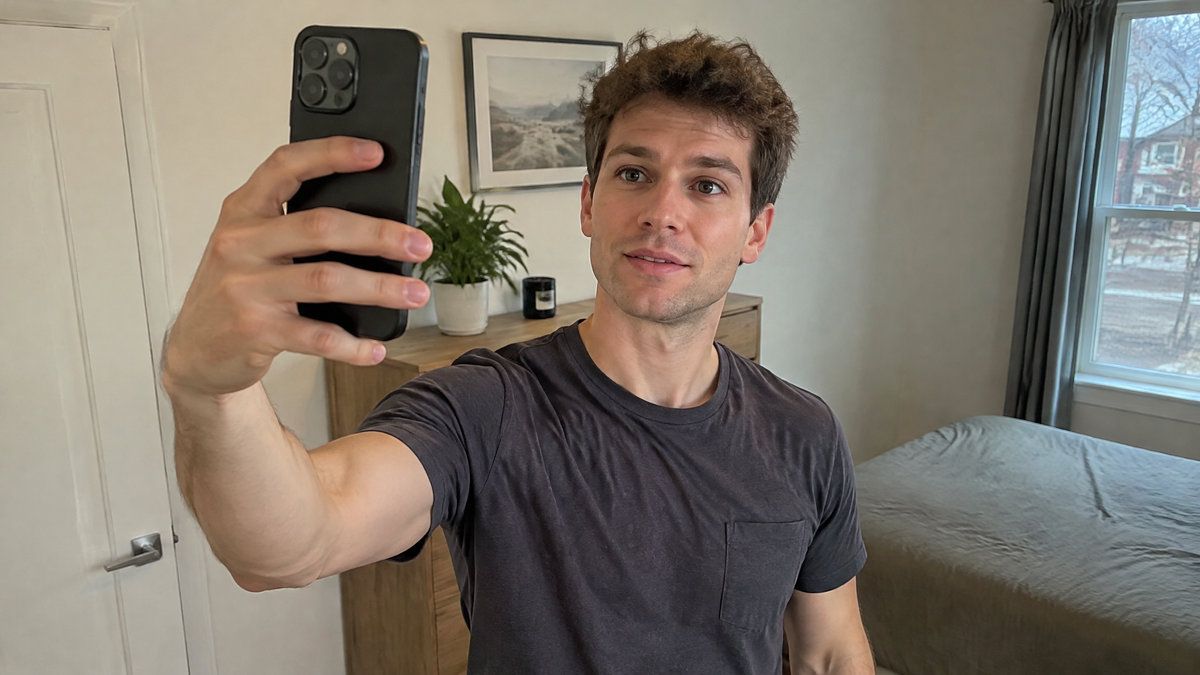
Capture an Accurate Baseline: Take a sharply focused photograph under natural daylight, holding the camera perpendicular to the skin, with a ruler or coin in the frame. Record the date and the exact anatomical location.
-
Check Against the "Red Flags": Arrange an in-person assessment promptly if a lesion exhibits irregular or notched borders, varies in colour, exceeds 6 mm in diameter, bleeds, crusts, causes persistent pain, or stands out as noticeably different from all your other spots (the "ugly duckling" sign).
-
Factor in Personal Vulnerability: Maintain a lower threshold for seeking medical review if you have a family history of skin cancer, numerous atypical moles, significant sun exposure, or a compromised immune system.
-
Treat Images as a Bridge, Not a Destination: Bring your photos to a GP or dermatologist to demonstrate how the lesion has evolved, but do not rely on them—or an automated algorithm—to decide whether care is necessary.
Where specialist access is limited, rely on qualified teledermatology services provided by hospital networks and licenced clinicians, rather than unsupervised diagnostic software. Use your smartphone to initiate an informed conversation with a medical professional, never to conclude it.